Facilities
Facility Statement
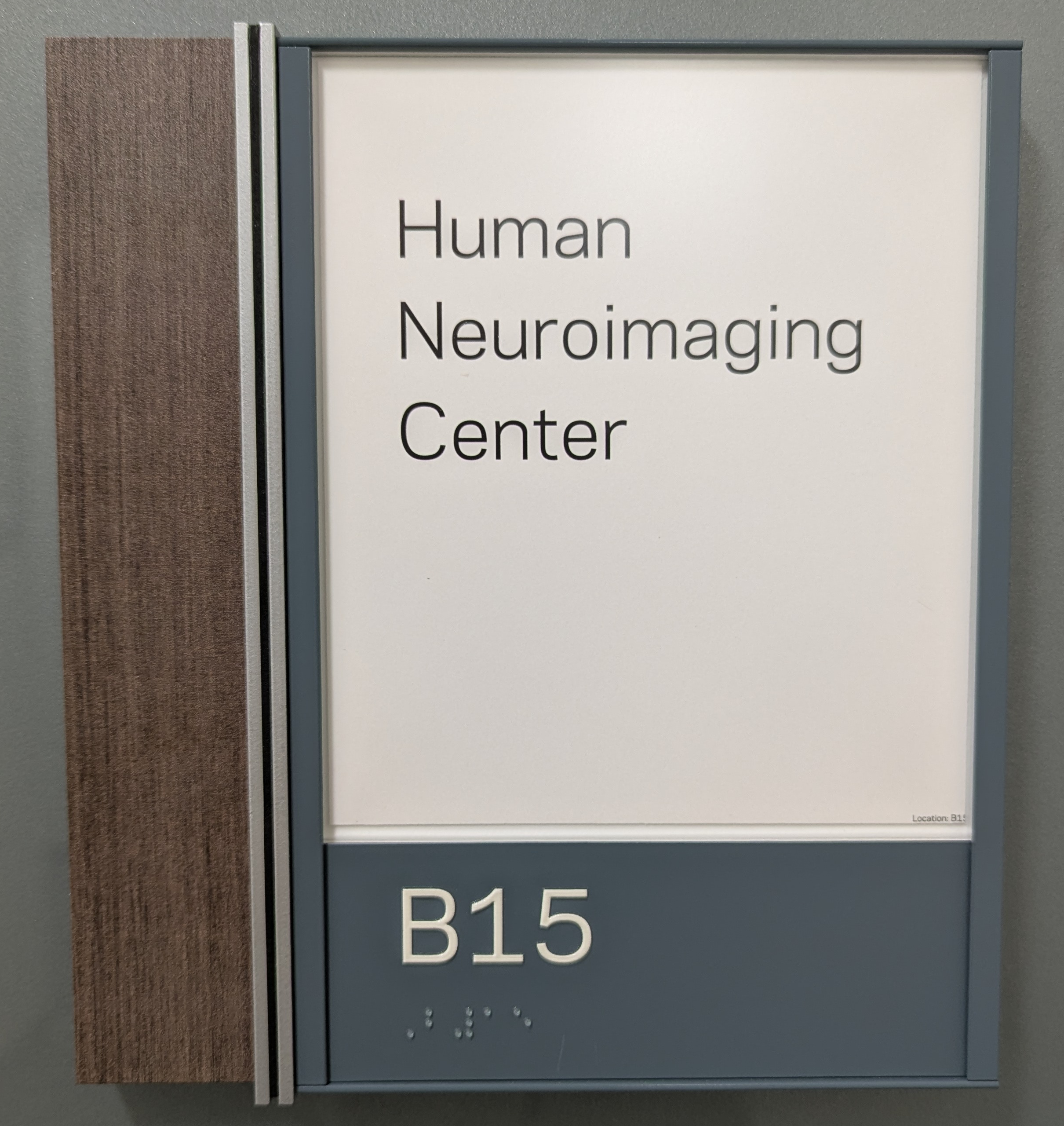
Notre Dame Human Neuroimaging Center
The Notre Dame Human Neuroimaging Center (ND-HNC) is located on the Lower Level of the Veldman Family Psychology Clinic, a dedicated 35,000-square-foot clinical and research facility located in South Bend, Indiana. The facility consists of a fully integrated environment for participant recruitment, clinical assessment, neuroimaging, electrophysiological recording, noninvasive brain stimulation, data processing, and collaborative scientific analysis.
The ND-HNC includes dedicated participant-facing spaces consisting of a reception area (117 ft\(^2\)), two consultation rooms (83 and 100 ft\(^2\)), two changing rooms (80 and 79 ft\(^2\)), and two interview rooms (110 and 112 ft\(^2\)). Research-dedicated spaces include a mock magnetic resonance imaging (MRI) scanner room (226 ft\(^2\)), two electroencephalogrphy (EEG) laboratories (157 and 161 ft\(^2\)), an EEG control room (104 ft\(^2\)), EEG preparation room (182 ft\(^2\)), MRI vestibule (308 ft\(^2\)), MRI storage room (179 ft\(^2\)), MRI scanner and control suite (~500 ft\(^2\)), a dedicated transcranial magnetic stimulation (TMS) laboratory (385 ft\(^2\)), image processing room (270 ft\(^2\)), conference room (257 ft\(^2\)), and three staff offices (121, 102, and 103 ft\(^2\)).
Magnetic Resonance Imaging Resources
Human neuroimaging studies are performed on a Siemens Healthineers MAGNETOM Cima.X 3 Tesla (3T) MRI system operating on the syngo MR XA61 platform. The system combines advanced research capabilities with a participant-friendly short-bore design featuring a 60 cm open bore architecture.
The scanner incorporates a highly homogeneous superconducting 3T magnet with exceptional field stability (\(\lt 0.1\) ppm/hour) and high-performance passive and active shimming capabilities. Automated subject-specific 3D shimming can be completed in approximately 15 seconds, ensuring optimal magnetic field homogeneity for advanced neuroimaging applications. Fifth-generation active shielding and Siemens External Interference Shielding (E.I.S.) technology minimize environmental magnetic disturbances and maintain image quality during demanding functional and diffusion imaging studies.
Image acquisition is supported by Siemens Gemini gradients capable of maximum gradient amplitudes of \(\ge 200\) mT/m per axis and slew rates of 200 T/m/s per axis, with vector performance reaching 346 mT/m and 346 T/m/s. These capabilities enable high-resolution structural imaging, advanced diffusion imaging, and rapid functional MRI acquisition with reduced distortion and improved signal fidelity.
The system employs Siemens DirectRF digital architecture and TimTX TrueForm/TrueShape parallel transmit technology to optimize radiofrequency (RF) homogeneity and signal stability. 204 coil elements and 64 simultaneous receiver channels can be utilized during data acquisition, providing high signal-to-noise ratios and supporting accelerated imaging protocols.
Advanced imaging capabilities include diffusion imaging with b-values up to 16,000 s/mm\(^2\), automated prospective motion correction with real-time six-degree-of-freedom tracking, parallel-transmit-enabled selective excitation (ZOOMit), and AI-assisted workflow tools through Siemens BioMatrix and myExam Companion technologies. These capabilities support state-of-the-art structural MRI, functional MRI, diffusion tensor imaging, tractography, and quantitative neuroimaging investigations.
Functional MRI Research Infrastructure
Functional MRI studies are supported by a comprehensive NordicNeuroLab (NNL) hardware and software ecosystem designed specifically for advanced neuroimaging research and clinical mapping applications.
Visual stimulus presentation is provided through a 40 in MR-compatible 4K UHD display system. These systems support high-resolution visual paradigms, visual field mapping, immersive task presentation, and real-time participant monitoring.
Behavioral responses are collected using MR-compatible fiber-optic ResponseGrips that minimize participant movement while providing reliable acquisition of task performance data. Synchronization between MRI pulse sequences and experimental paradigms is achieved through the NNL SyncBox system, which provides scanner-independent synchronization with timing accuracy of ±0.5 ms.
Experimental paradigm delivery is managed using nordicAktiva software, which includes validated libraries of motor, language, cognitive, and clinical paradigms while also supporting custom paradigm development. The platform permits synchronized control of stimulus presentation and image acquisition and provides rehearsal modes for participant training.
Advanced image processing is supported by nordicMEDiVA, a web-based neuroimaging analysis environment supporting task-based fMRI, diffusion imaging, tractography, and perfusion analysis. Automated preprocessing includes motion correction, spatial normalization, diffusion distortion correction, and statistical modeling using General Linear Models. Interactive tools support activation mapping, tract reconstruction, region-of-interest (ROI) based analyses, and collaborative review among multiple investigators.
Simulation and Participant Training Resources
The facility houses an Encore MRI Simulator (Psychology Software Tools/RadSim), which provides a realistic MRI environment for participant acclimation, protocol piloting, and training. The simulator includes a realistic 60 cm bore, motorized participant table, integrated positioning lasers, environmental lighting, cooling systems, and participant safety monitoring.
The simulator reproduces scanner acoustics and vibration using high-fidelity recordings obtained from Siemens MRI systems. Visual presentation is delivered through a dedicated 22 in high-definition display with integrated audio communication and real-time participant monitoring through an in-bore camera system.
The simulator incorporates interchangeable mock head coils replicating Siemens acquisition environments and includes response devices for behavioral task practice. The MoTrak motion tracking system provides real-time monitoring of participant head motion across three translational and three rotational axes, allowing participants to receive immediate feedback regarding movement. This capability substantially improves participant compliance and reduces motion-related data loss during actual MRI sessions.
Research Personnel and Technical Support
To ensure the highest standards of data quality, regulatory compliance, and operational efficiency, the Notre Dame Human Neuroimaging Center provides dedicated, expert human infrastructure. The facility is staffed by a specialized team available to support investigators throughout the lifecycle of their research projects
Director: The senior scientist/faculty member provides overall strategic vision, scientific leadership, and governance for the neuroimaging center. The Director establishes facility policies, reviews incoming research protocols for scientific merit and technical feasibility, fosters interdisciplinary collaborations, and ensures the center’s capabilities remain at the cutting edge of neuroimaging technology.
Scientific Program Coordinator: The Coordinator facilitates the center’s broader academic and research mission. Key responsibilities include managing the center’s pilot grant funding programs, organizing educational workshops and scanner user seminars, assisting investigators with grant submission logistics, and coordinating internal and external scientific communications and progress reporting.
Physicist: The physicist is dedicated to the center to oversee technical operations, optimize imaging sequences, and develop custom acquisition protocols tailored to specific research questions. The Physicist manages the system’s rigorous quality assurance (QA) program: continually tracking signal-to-noise ratios (SNR), field stability, and artifact management. The Physicist also provides advanced consultation on experimental design and safety guidelines.
Analyst: The neuroimaging data expert provides specialized support for advanced image processing, pipeline development, and computational workflows. The Analyst maintains the center’s containerized processing pipelines (e.g., fMRIPrep, FreeSurfer, FSL, AFNI) across local workstations and the University’s High-Performance Computing (HPC) cluster, assisting investigators with data curation, quality control (QC) metrics, and reproducible statistical modeling. They Analyst also provides bootcamps and courses covering topics from programming to modelling multimodal MRI data, making the incorporation of MRI techniques into existing research programs more accessible.
Technologist: A certified, full-time Technologist operates the 3T scanner and handles daily scanning execution. The Technologist is responsible for strict participant safety screening, anatomical positioning, and the seamless integration of peripheral equipment (including the NordicNeuroLab stimulus presentation systems and response devices). They ensure comfortable, safe, and highly standardized data collection environments for all participant cohorts.
Operations Coordinator: The Coordinator manages the day-to-day administrative and logistical workflows of the facility. Key responsibilities include administering the centralized participant scheduling system, coordinating billing, ensuring compliance with institutional IRB and federal regulations, and facilitating investigator onboarding and safety training. The Coordinator serves as the primary operational liaison between research teams and the center.
Electrophysiology and Neuromodulation Resources
The facility contains two dedicated EEG acquisition laboratories, a centralized EEG control room, and a participant preparation suite supporting high-density electrophysiological recording. These resources facilitate cognitive, developmental, affective, and clinical neuroscience investigations requiring millisecond temporal resolution.
Noninvasive brain stimulation studies are conducted in a dedicated TMS laboratory equipped for experimental and translational neuromodulation research. The co-location of MRI, EEG, and TMS resources supports multimodal neuroimaging and neurostimulation investigations.
Data Processing, Storage, and Computational Resources
Imaging Workstations and Local Data Infrastructure
Primary image management and analysis are supported by Apple Mac Studio workstations utilizing Apple Silicon architectures with unified memory systems, hardware-accelerated media processing, and dedicated neural processing engines. These systems support image visualization, machine learning workflows, statistical analysis, and neuroimaging software development.
The workstations are connected to a dedicated Promise Technology Pegasus RAID storage subsystem providing 104 TB of fault-tolerant local storage. The storage architecture utilizes hardware RAID redundancy and predictive data migration technologies to ensure data integrity and uninterrupted operation. High-bandwidth Thunderbolt connectivity supports efficient processing of large multimodal imaging datasets.
DICOM image management, visualization, and anonymization are performed using the Horos imaging platform. Investigators have access to advanced multiplanar reconstruction, volume rendering, quantitative ROI analysis, and secure de-identification tools that support compliance with institutional and federal human subjects protections.
Departmental Computing Resources
The research team maintains access to a dedicated departmental computing rack, providing 56 CPU cores and 256 GB RAM for local high-throughput processing. This infrastructure supports image reconstruction, quality assurance, machine learning development, containerized neuroimaging pipelines, and rapid deployment of computational workflows.
Dedicated Linux workstations provide optimized environments for FSL, FreeSurfer, AFNI, ANTs, and other neuroimaging software packages. These systems include dedicated GPU acceleration to support computationally intensive analyses such as probabilistic tractography, deep-learning-based segmentation, and large-scale image processing.
Institutional High-Performance Computing Resources
The project additionally has access to the University’s High-Performance Computing (HPC) infrastructure through the Center for Research Computing. This resource provides approximately 1,280 dedicated MRI-analysis CPU cores interconnected through a high-bandwidth, low-latency network fabric designed for parallel scientific computing.
The HPC environment is integrated with approximately 800 terabytes of enterprise-grade storage capable of supporting large-scale neuroimaging studies, longitudinal datasets, and multimodal analyses. Advanced workload management systems facilitate efficient execution of computationally intensive pipelines including fMRIPrep, FreeSurfer, diffusion tractography, machine learning workflows, and group-level statistical analyses.
Overall Research Environment
The Veldman Family Psychology Clinic and Notre Dame Human Neuroimaging Center provide an exceptional environment for NIH-funded neuroscience and behavioral research. The co-location of advanced MRI, EEG, TMS, participant training facilities, dedicated technical personnel, extensive computational infrastructure, and large-scale data storage resources enables efficient implementation of sophisticated multimodal research protocols. These integrated facilities provide all equipment, personnel, and computational resources necessary to successfully conduct the proposed studies and support high-quality, reproducible neuroimaging research.